Open Access | Review
This work is licensed under a Creative Commons Attribution-ShareAlike 4.0 International License.
Resilience in the depths: anemones as models for aging and regeneration research
* Corresponding author: Pooja B. Rasal
Mailing address: Department of Pharmacology, JES's SND College of Pharmacy, Babhulgaon (Yeola), India.
Email: poojarasal2000@gmail.com
Received: 25 April 2025 / Revised: 23 May 2025 / Accepted: 20 June 2025 / Published: 29 September 2025
DOI: 10.31491/APT.2025.09.179
Abstract
All creatures are impacted by the complicated biological process of aging, although little is known about its exact mechanics. Because of their simple body shape and amazing capacity for regeneration, sea anemones, which belong to the phylum Cnidaria, provide a unique model for researching aging. With an emphasis on their cellular and molecular traits, capacity for regeneration, and environmental effects, this article examines the biology of anemones in relation to aging. The anatomy and physiology of anemones are first described, with a focus on the function of specific stem cells in regeneration. Senescence and decreased stem cell activity are two major biological changes that these creatures undergo as they age and have an effect on their ability to regenerate. The life cycles of different anemone species and the variables affecting their lifespan are also covered. To clarify their roles in cellular aging, important molecular mechanisms of aging are investigated, including oxidative stress, genetic factors, and signaling pathways (such as Wnt, Notch, and Hedgehog). The aging process is also significantly influenced by environmental factors, such as pollution and temperature. There are also issues, such as the paucity of longitudinal studies, despite their usefulness as models for aging research. But new methods in imaging and molecular biology are starting to fill up these gaps. To sum up, research on anemone aging advances our knowledge of their biology and has wider ramifications for aging studies in other animals and regenerative medicine. We may discover a great deal about the basic mechanisms governing lifespan and resilience in living things by investigating the interactions between aging, regeneration, and environmental conditions.
Keywords
Aging, sea anemones, regeneration, longevity
Introduction
Aging, or senescence, is a complex biological process marked by the progressive decline of physiological functions and increased susceptibility to disease in multicellular organisms. This process is driven by a range of genetic, environmental, and lifestyle factors, leading to cellular and molecular changes such as protein damage accumulation, mitochondrial dysfunction, telomere shortening, and impaired intercellular communication. These alterations collectively contribute to tissue degeneration, cellular senescence, and systemic dysfunctions that influence both longevity and overall health [1]. Beyond its biological implications, aging has far-reaching social and economic consequences. The rising incidence of age-related diseases including cancer, cardiovascular disorders, and neurodegenerative conditions places increasing pressure on global healthcare systems. As a result, there is an urgent need to understand the underlying mechanisms of aging in order to develop effective interventions that promote healthy aging and extend lifespan [2]. Central to this endeavor is the study of stem cells, which are critical for tissue maintenance and regeneration. With age, stem cell function declines, manifested through reduced proliferation, altered differentiation capacity, and increased cellular senescence. These impairments compromise the ability of tissues to repair damage and maintain homeostasis [2].
Recent studies have shown that enhancing stem cell activity or mitigating the factors contributing to their decline can improve tissue regeneration and delay the onset of aging symptoms. Experimental treatments that stimulate stem cell function have yielded promising results in model organisms, such as fruit flies and mice, suggesting potential therapeutic applications for age-related conditions [3, 4]. This study explores the molecular and cellular mechanisms of aging by utilizing Nematostella vectensis, a basal metazoan from the phylum Cnidaria, as a model organism. N. vectensis offers unique advantages for aging and regenerative research due to its simple body plan, remarkable regenerative capabilities, and well-characterized genome. Importantly, it possesses diverse stem cell populations that actively contribute to tissue renewal, making it an ideal system for investigating stem cell dynamics across the lifespan. Furthermore, its phylogenetic position allows researchers to explore the evolutionary conservation of aging and regeneration mechanisms across metazoans [5, 6].
The main objective of this study is to characterize how stem cell function in N. vectensis changes with age and under environmental stress, and to identify molecular pathways involved in these processes. By analyzing gene expression patterns, cellular turnover rates, and regenerative capacity in aging polyps, this research aims to uncover key factors that regulate stem cell activity and longevity. Insights gained from N. vectensis not only deepen our understanding of fundamental aging biology but may also inform strategies for improving stem cell function and regenerative therapies in more complex organisms, including humans [7].
Biology of anemone stem cells
Because of their extraordinary capacity for regeneration and straightforward body structure, anemones especially sea anemones belonging to the phylum Cnidaria make intriguing subjects for stem cell research.
Basic structure of anemones
The cylindrical body of an anemone has a central mouth that is encircled by tentacles (Figure 1). The mesoglea, a gelatinous substance, sits between the outer epidermis and the inner gastrodermis, which make up their two primary layers. Instead of having a centralized nervous system, they have a rudimentary nerve net that enables simple reactions to stimuli [8].
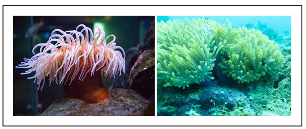
Figure 1. Sea Anemone.
Types of stem cells in anemones
An unusual collection of stem cells seen in anemones, especially sea anemones, helps explain their extraordinary capacity for regeneration. In order to preserve their cellular diversity and carry out amazing regenerative tasks, anemones rely on a specific population of interstitial stem cells [9]. Their resilience and flexibility in a variety of settings are largely due to their capacity to transition between stem and differentiated states. Different types for stem cell anemone are mentioned in Table 1.
Table 1.
Types of stem cells in anemones.
| Stem cell Type | Location | Function | Characteristics | Ref. |
|---|---|---|---|---|
| Epithelial stem cells | Epidermis and gastrodermis | Contribute to epithelial turnover and regeneration | High proliferation rate, multipotent | [10] |
| Mesenchymal stem cells | Mesoglea | Differentiate into muscle, nerve, and other cell types | Can migrate, multipotent | [11] |
| Nerve stem cells | Nerve net | Regenerate nerve cells after injury | Exhibit neural differentiation potential | [8] |
| Germline stem cells | Gonads | Responsible for gametogenesis | Can give rise to sperm and eggs, regulated by environmental cues | [12] |
| Planula stem cells | Developing larvae (planula stage) | Enable metamorphosis and tissue formation | Unique to developmental stages, high plasticity | [13] |
| Ciliated stem cells | Epidermal layer | Contribute to ciliary regeneration | Highly proliferative, involved in sensory functions | [14] |
| Muscle stem cells | Mesoglea and muscle tissue | Regenerate muscle fibers | Can differentiate into various muscle types | [6] |
| Epithelial-mesenchymal transition (EMT) cells | Various tissues | Facilitate wound healing and regeneration | Plasticity, can switch between epithelial and mesenchymal states | [15] |
| Stolon stem cells | Stolon regions (in colonial species) | Enable asexual reproduction and growth | Multipotent, involved in forming new polyps | [16] |
Mechanism of stem cell renewal and differentiation
The regulation of stem cell renewal and differentiation in sea anemones, such as Nematostella vectensis, involves molecular pathways that are not only conserved across metazoans but also exhibit unique adaptations. These pathways are integral to the anemone's remarkable regenerative abilities and tissue homeostasis. Below are the key pathways studied in the context of anemones:
Wnt/β-catenin pathway
The Wnt/β-catenin signaling pathway is a fundamental molecular mechanism that governs numerous developmental and regenerative processes in many multicellular organisms, including the sea anemone Nematostella vectensis. In Nematostella, this pathway is crucial for axial patterning, specifically the establishment and maintenance of the oral-aboral axis, which is essential for organizing body structure and polarity during both embryonic development and regeneration. One of the central components of this pathway is β-catenin, a protein that, when stabilized by Wnt ligands, accumulates in the cytoplasm and translocates into the nucleus. Once in the nucleus, β-catenin acts as a transcriptional co-activator, partnering with TCF/LEF transcription factors to activate gene expression programs that sustain stem cell pluripotency and proliferation. In Nematostella, Wnt/β-catenin signaling is especially active in areas undergoing regeneration, such as around wounds or at the oral pole during body part regrowth. This elevated signaling promotes cellular proliferation and differentiation, facilitating the replacement of lost or damaged tissues. Notably, experimental studies have shown that interference with Wnt signaling, either through genetic manipulation or pharmacological inhibition, can severely impair regenerative outcomes. Disruption of this pathway leads to loss of axial patterning, misexpression of key developmental genes, and a failure to maintain stem cell populations, ultimately resulting in compromised tissue integrity. These findings emphasize the central role of Wnt/β-catenin signaling in governing stem cell behavior, orchestrating regeneration, and preserving the overall structural organization in Nematostella vectensis. Because of its evolutionary conservation, insights gained from studying this pathway in anemones can inform broader understanding of regeneration and stem cell biology in more complex animals, including humans [17].
Notch signaling pathway
The Notch signaling pathway is a highly conserved cell communication mechanism that plays a crucial role in cell fate determination, tissue development, and regeneration across metazoans. In the cnidarian Nematostella vectensis, Notch signaling is particularly vital for regulating the behavior of interstitial stem cells—a multipotent stem cell population responsible for generating diverse cell types, including nematocytes (stinging cells) and neurons. This pathway enables cells to make tightly regulated decisions about whether to remain in a stem-like state or differentiate into specialized lineages, thus maintaining tissue homeostasis and proper developmental patterning. In Nematostella, Notch signaling operates largely through a mechanism known as lateral inhibition, where interactions between neighboring cells influence their developmental fates. When Notch receptors on one cell interact with ligands (such as Delta or Jagged) on an adjacent cell, a cascade is initiated that leads to the cleavage of the Notch receptor. The cleaved intracellular domain (NICD) translocates to the nucleus, where it activates target genes that suppress differentiation pathways, thereby maintaining the stemness of the receiving cell. This system ensures that not all neighboring cells differentiate at the same time, creating a balance between stem cells and differentiated cells during development and especially during regenerative processes or tissue remodeling.
Experimental disruption of Notch signaling in Nematostella such as through gene knockdown or chemical inhibitors has demonstrated the critical nature of this pathway. Loss of Notch activity results in the overproduction of differentiated cells at the expense of the stem cell pool, ultimately compromising the organism's ability to regenerate tissues effectively. Conversely, overactivation can suppress necessary differentiation, leading to an accumulation of undifferentiated cells and impaired tissue structure. These findings underscore Notch signaling as a regulatory checkpoint that preserves the delicate equilibrium between proliferation and differentiation, making it a central component in the anemone’s regenerative toolkit. The insights from Nematostella's Notch pathway not only illuminate ancient mechanisms of developmental biology but also provide valuable parallels for understanding stem cell regulation and regenerative medicine in higher organisms [18].
BMP (bone morphogenetic protein) signaling
The BMP (bone morphogenetic protein) signaling pathway is a key regulator of embryonic development and tissue differentiation in many animals, and in Nematostella vectensis, it plays a vital role in establishing and maintaining germ layer identities—particularly the ectoderm and endoderm. BMPs are members of the TGF-β (transforming growth factor-beta) superfamily and function through a highly conserved mechanism in which BMP ligands bind to serine/threonine kinase receptors on the cell surface. This leads to phosphorylation of receptor-regulated SMAD proteins, which then translocate into the nucleus to influence the expression of genes involved in cell fate specification and tissue patterning. In Nematostella, BMP signaling is especially important during early development, where it contributes to defining the dorsoventral axis and helps demarcate tissue boundaries. Beyond initial development, BMP signaling continues to play a role in tissue maintenance and regeneration. It has been shown to regulate the differentiation of pluripotent stem cells into specific germ layer derivatives by providing context-specific cues that guide cells toward ectodermal or endodermal fates. Importantly, BMP signaling does not act in isolation; it functions in conjunction with the Wnt/β-catenin pathway, forming a complex signaling network that modulates both stem cell maintenance and lineage commitment. This interplay ensures that a precise number of cells differentiate while a sufficient pool of pluripotent cells is retained, which is essential for successful regeneration and proper tissue remodeling.
Experimental studies in Nematostella have demonstrated that perturbation of BMP signaling, either through genetic interference or molecular inhibitors, disrupts the normal patterning of tissues, resulting in abnormal germ layer formation and impaired regenerative capabilities. For example, inhibition of BMP signaling during regeneration can lead to a failure in the re-establishment of organized tissue layers, highlighting its critical role not just in embryogenesis but also in adult tissue renewal. These findings underscore BMP signaling as a developmental and regenerative linchpin, offering insights into how ancient regulatory pathways control stem cell differentiation and tissue architecture. Moreover, the conservation of BMP function across species suggests that understanding its role in Nematostella may provide valuable clues for enhancing stem cell-based therapies and regenerative medicine in humans. [21].
Hedgehog signaling pathway
The Hedgehog (Hh) signaling pathway is a crucial regulator of developmental processes and tissue regeneration, and in sea anemones, such as Nematostella vectensis, it plays an important role in tissue patterning, stem cell proliferation, and regenerative growth. Hedgehog signaling is known for its role in transmitting information required for the proper spatial arrangement of tissues during embryogenesis, but in sea anemones, it extends its function into post-embryonic regeneration, supporting the organism's remarkable ability to heal and regrow damaged or lost body parts. The pathway operates through the interaction of Hedgehog ligands with the Patched (PTCH) receptor. In the absence of a Hedgehog signal, PTCH inhibits the activity of another membrane protein called Smoothened (SMO). When Hedgehog ligands are present, they bind to PTCH, relieving this inhibition and allowing SMO to activate downstream intracellular components that eventually regulate Gli transcription factors. These factors then enter the nucleus and control the expression of genes involved in cell proliferation, differentiation, and pattern formation.
In Nematostella, research has identified Hedgehog signaling activity in regions undergoing active regeneration, such as near wounds or amputation sites. Here, the pathway contributes to the re-establishment of tissue architecture by guiding stem cells toward appropriate fates and promoting organized proliferation. Studies have shown that disruption of Hedgehog signaling can result in defective regeneration, including disorganized tissue growth and incomplete recovery of body structures. This highlights the pathway’s essential role in coordinating not only when and where cells divide, but also how they differentiate to restore functionally integrated tissues. Furthermore, Hedgehog signaling often acts in concert with other key developmental pathways, such as Wnt and BMP, forming a complex signaling network that ensures precise spatial and temporal control of gene expression during regeneration. The ability of Nematostella to use Hedgehog signaling for such sophisticated regenerative purposes underscores its evolutionary significance and suggests that deeper understanding of this pathway in simple organisms can provide profound insights into regenerative biology, stem cell regulation, and potential therapeutic targets in more complex animals, including humans [19].
PI3K/AKT pathway
The PI3K/AKT signaling pathway is a central regulator of cell survival, metabolism, and proliferation, and in Nematostella vectensis, it plays a crucial role in supporting the survival and self-renewal of stem cells, particularly during regeneration and tissue maintenance. This pathway is activated when phosphoinositide 3-kinases (PI3Ks) respond to extracellular signals—such as growth factors or cytokines—by phosphorylating lipid messengers in the cell membrane. These lipid messengers recruit and activate AKT (also known as Protein Kinase B), which then phosphorylates a variety of downstream targets to regulate cellular processes essential for regeneration. In Nematostella, activation of the PI3K/AKT pathway promotes cell cycle progression, enabling stem cells to proliferate in response to injury or during routine tissue renewal. It also plays a critical role in inhibiting apoptosis (programmed cell death), ensuring that stem cell populations remain viable and capable of sustained activity. This is particularly important during regeneration, when stem cells must not only survive in a potentially hostile or damaged environment but also expand in number to replace lost or damaged tissues. Additionally, PI3K/AKT signaling supports metabolic activity, helping stem cells meet the high energy demands associated with rapid proliferation and differentiation.
Experimental findings in Nematostella suggest that impairing this pathway—through inhibition of PI3K or AKT—leads to a marked reduction in cell proliferation, increased cell death, and compromised regenerative outcomes. Conversely, enhanced PI3K/AKT activity correlates with increased stem cell robustness and more efficient regeneration. Moreover, the PI3K/AKT pathway often interacts with other signaling cascades, such as the Wnt and Notch pathways, forming an integrated network that finely tunes stem cell behavior and tissue architecture. The significance of the PI3K/AKT pathway in Nematostella mirrors its vital functions in higher organisms, including humans, where it is also linked to stem cell maintenance, cancer biology, and age-related decline. Understanding how this pathway operates in a simple yet highly regenerative organism like Nematostella offers valuable insight into how similar mechanisms might be harnessed or modulated in human regenerative medicine, anti-aging strategies, and stem cell therapies [20].
Role of stem cells in anemone aging
While stem cell dysfunction is well-documented in humans and other vertebrates, many fundamental questions about how aging affects stem cell biology remain unresolved. To address these questions, researchers are increasingly turning to simpler model organisms that offer unique insights into conserved biological processes. In this context, the sea anemone Nematostella vectensis serves as a valuable model due to its evolutionary position and remarkable regenerative capabilities. Studying such basal metazoans provides an opportunity to uncover the core principles governing stem cell maintenance and aging across species.
Stem cells play a critical role in the aging process of sea anemones, especially in species like Nematostella vectensis. These organisms demonstrate an exceptional ability to regenerate and maintain tissue integrity, which is largely attributed to the constant activity of their stem cells. Their regenerative abilities and the absence of typical signs of aging, such as senescence, have made sea anemones a focal point in studies of longevity and regenerative medicine. Stem cells in anemones contribute significantly to cellular upkeep, stress resistance, and the continuous renewal of tissues, making them central to the organism's maintenance of youthful vigor over long periods. Below are keys aspects of stem cell function in anemone aging, supported by evidence from recent studies.
Continuous regeneration
Sea anemones are equipped with pluripotent stem cells, which enable them to regenerate lost body parts, such as tentacles and damaged tissues. These stem cells can differentiate into a variety of specialized cell types, including muscle, neuron, and epithelial cells, to restore damaged tissues or even entire body structures. The regenerative capacity of sea anemones is remarkable in its ability to adapt to environmental changes, such as temperature fluctuations and mechanical damage. Complex signaling pathways such as Wnt/β-catenin, Notch, and Hedgehog pathways regulate this continuous regeneration, ensuring that the cells involved in regeneration are properly activated, differentiated, and integrated into the existing tissue structures. Research on these pathways in sea anemones provides valuable insights into the molecular mechanisms of regeneration, which could have significant implications for understanding aging in other species [10, 21].
Stem cell maintenance and homeostasis
The long-term health and regenerative potential of sea anemones depend heavily on the continuous maintenance of their stem cell populations. These cells undergo regular divisions to produce new progeny that replace aged or damaged cells, thus contributing to the organism's ability to resist the degenerative effects of aging. This regenerative process is not just a response to injury but is continuously active, ensuring that cellular homeostasis is preserved throughout the anemone's life. For instance, Nematostella vectensis possesses a specialized population of stem cells that are maintained through dynamic regulation by genes such as nanos and piwi, which are crucial for maintaining stem cell pluripotency and preventing differentiation. The efficient management of stem cell renewal ensures the preservation of tissue integrity and function, slowing down the appearance of age-related degenerative changes [21]. This continuous turnover and renewal of cells help to sustain the organism's overall fitness and adaptability across various environmental conditions [22].
Response to environmental stress
Sea anemones are highly adaptable to fluctuating environmental conditions, such as changes in temperature, salinity, and the presence of pollutants. Their stem cells play a key role in this environmental adaptability by responding to stress and minimizing cellular damage. Upon exposure to stressful conditions, sea anemones activate stress response pathways that protect cellular components from damage. Heat shock proteins (HSPs) and antioxidant systems are upregulated to manage protein folding and reduce oxidative stress. These mechanisms promote cellular repair and recovery, which contribute to the organism's overall longevity and regenerative abilities. The ability of sea anemones to maintain stem cell activity under stress conditions supports the notion that these organisms can survive in harsh environments over extended periods, further enhancing their capacity for longevity [10, 22].
Minimal cellular senescence
Unlike many other organisms, sea anemones exhibit minimal evidence of cellular senescence as they age. Cellular senescence refers to a state in which cells permanently stop dividing, contributing to tissue dysfunction and aging. In contrast, anemones maintain active stem cell populations that continue to support tissue growth and repair throughout the organism's lifespan. Research suggests that sea anemones have developed specialized mechanisms to preserve their stem cells, potentially through enhanced DNA repair pathways and mechanisms that protect telomere length. Telomeres, the protective caps at the ends of chromosomes, play a key role in cellular aging. In Nematostella vectensis, telomeres appear to be maintained or repaired over time, preventing the accumulation of senescent cells and supporting the ongoing function of stem cells. This ability to avoid senescence is a significant factor in the organism's ability to resist the typical aging processes seen in other species [23].
Genetic and molecular pathways
Critical genetic and molecular pathways, such as Wnt, Notch, and Hedgehog signaling, regulate the maintenance and differentiation of stem cells in sea anemones. These pathways are highly conserved across species and play pivotal roles in cellular development, tissue regeneration, and homeostasis. The Wnt/β-catenin pathway is involved in stem cell self-renewal and differentiation, while Notch signaling is crucial for maintaining the balance between stem cell maintenance and differentiation into specialized cell types. Hedgehog signaling, on the other hand, controls tissue patterning and cell division during regeneration. Understanding how these pathways function in sea anemones can provide crucial insights into how stem cells contribute to longevity and regeneration in these organisms. Furthermore, these findings could have broader implications for aging research and regenerative medicine in other species, potentially identifying therapeutic targets for promoting tissue repair and longevity [21, 23].
Aging in anemones
Sea anemones, particularly Nematostella vectensis, are extraordinary organisms that exhibit negligible senescence and remarkable regenerative abilities. Their ability to maintain tissue homeostasis and avoid age-related decline provides valuable insights into aging and longevity. Molecular markers of aging, including telomere shortening, accumulation of senescence-associated beta-galactosidase (SA-β-gal), and activation of cell cycle inhibitors such as p16INK4a and p21, are largely absent or minimally expressed in anemones (Table 2). These molecular signatures, which are commonly associated with cellular aging in other organisms, are significantly less pronounced or non-existent in these organisms, suggesting that they have evolved mechanisms to resist cellular aging. Additionally, the activation of the Wnt and Notch signaling pathways in anemones plays a crucial role in promoting cell division, maintaining stem cell populations, and preserving the regenerative capacity throughout their lifespan. The ability of anemones to resist typical aging markers while sustaining regeneration raises intriguing questions about the evolutionary advantages of their regenerative capabilities. This phenomenon could provide valuable insights into tissue repair, longevity, and age-related diseases in other species, offering potential avenues for developing therapeutic strategies that delay or reverse aging processes 6, 24]. Below, the biological changes associated with aging in sea anemones (Figure 2).
Protein misfolding and aggregation
Protein homeostasis, or proteostasis, plays a vital role in preventing cellular damage and maintaining health in sea anemones over time. Like other organisms, sea anemones experience protein misfolding, which can lead to the accumulation of misfolded proteins and interfere with cellular functions if not managed properly. However, sea anemones possess a robust system to mitigate these effects. Heat shock proteins (HSPs), a group of molecular chaperones, are particularly crucial in stabilizing misfolded proteins and facilitating their refolding or degradation. This effective maintenance of proteostasis enables sea anemones to avoid the cellular stress and damage that contribute to aging in other species, where the accumulation of misfolded proteins is often linked to diseases such as Alzheimer’s and Parkinson’s [23].
Stem cell dynamics
Sea anemones exhibit remarkable tissue regeneration capabilities, largely due to their populations of multipotent stem cells. These stem cells are regulated by evolutionarily conserved genes like nanos and piwi. Unlike other organisms, where these genes are primarily associated with germline maintenance, in sea anemones, they ensure the long-term activity of somatic stem cells. These genes help sustain stem cell potency and enable continuous differentiation into various cell types required for tissue repair and regeneration. Unlike aging organisms where stem cell exhaustion leads to reduced regenerative capacity, sea anemones maintain active and potent stem cell populations throughout their lives, contributing significantly to their negligible senescence and exceptional longevity [21].
Regenerative capacity and tissue remodeling
One of the most distinctive traits of sea anemones is their extraordinary regenerative capacity, which allows them to replace lost or damaged tissues efficiently, even after severe injuries. This process is driven by the activation of their stem cell populations, which proliferate rapidly and migrate to the injury site. During regeneration, critical signaling pathways such as Wnt/β-catenin, Notch, and Hedgehog are upregulated, orchestrating cell division, differentiation, and tissue patterning. Morphogenetic gradients further guide tissue remodeling, ensuring that regenerated tissues are functional and correctly patterned. Beyond injury response, sea anemones also undergo continuous tissue turnover, a process that helps maintain cellular integrity and combat the effects of aging over time [10].
Absence of tumor formation
Despite their high rates of cellular proliferation, sea anemones rarely develop tumors, which is a significant factor in their sustained health and longevity. This rarity is attributed to tightly regulated mechanisms that control cell division and prevent unchecked growth. Cell cycle checkpoints ensure proper cell division, while programmed cell death, or apoptosis, removes cells with DNA damage or aberrant behavior. Additionally, sea anemones may possess innate immune-like responses capable of recognizing and eliminating potentially cancerous cells. These mechanisms collectively prevent tumorigenesis, a common hallmark of aging in other organisms, and further contribute to the longevity and health of sea anemones throughout their lives [22].
Table 2.
Molecular markers of aging.
| Molecular marker | Description | Function | Ref. |
|---|---|---|---|
| Telomere length | Shortening of telomeres with age, indicating cellular aging | Protects chromosome ends; critical for cell division | [25] |
| Senescence-associated secretory phenotype (SASP) | Release of pro-inflammatory cytokines by senescent cells | Contributes to tissue inflammation and aging | [26] |
| p16INK4a expression | Increased expression of this cyclin-dependent kinase inhibitor with age | Regulates cell cycle and inhibits proliferation | [27] |
| Mitochondrial dysfunction | Accumulation of mutations in mitochondrial DNA and decreased function | Impairs energy production and increases oxidative stress | [28] |
| Accumulation of advanced glycation end products (AGEs) | Increased AGEs contribute to tissue damage and inflammation | Alters protein structure and function; linked to chronic diseases | [29] |
| Changes in DNA methylation | Age-related alterations in DNA methylation patterns. | Affects gene expression and cellular function | [30] |
| Increased reactive oxygen species (ROS) | Higher levels of ROS contribute to oxidative stress | Causes damage to lipids, proteins, and DNA, accelerating aging | [31] |
| Loss of proteostasis | Impairment in the maintenance of protein homeostasis | Leads to aggregation of misfolded proteins, affecting cell function | [32] |
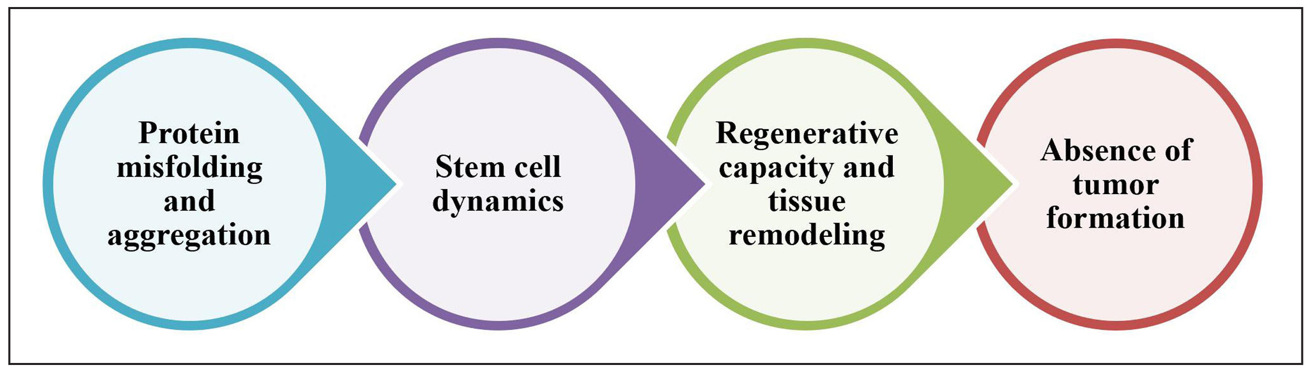
Figure 2. Biological changes associated with aging in sea anemones.
Comparison of aging process in anemone with other animals
The diversity of aging mechanisms across species is vividly illustrated through the comparison of organisms such as anemones, humans, mice, and Caenorhabditis elegans (C. elegans), as outlined in Table 3. Humans and mice represent the more familiar pattern of aging, characterized by progressive physiological decline, increased vulnerability to disease, and reduced regenerative capacity as they age. These organisms provide essential models for studying age-related diseases and understanding the cellular and systemic deterioration that occurs over time. In stark contrast, anemones exhibit remarkable regenerative abilities and show minimal signs of aging, challenging traditional definitions of senescence. Their biological resilience suggests the presence of unique molecular mechanisms that could potentially be harnessed for therapeutic regeneration and age-delaying strategies in humans. Meanwhile, C. elegans, a small nematode, serves as a powerful genetic model for aging research. Its short lifespan and well-mapped genome make it ideal for identifying specific genes and pathways that influence longevity. Research in C. elegans has led to the discovery of conserved genetic pathways, such as insulin/IGF-1 signaling, which are also relevant to aging in higher organisms. Collectively, the study of these diverse species not only enhances our fundamental understanding of aging but also opens promising avenues for advancements in regenerative medicine, age-related disease prevention, and longevity interventions.
Building on this comparative perspective, the regenerative capacity observed in anemones is particularly fascinating for scientists studying cellular immortality and tissue renewal. Unlike mammals, anemones can replace damaged tissues and even regenerate entire body parts with minimal decline in function over time. This ability is largely attributed to their abundant stem cells and efficient cellular repair mechanisms. These features allow anemones to maintain homeostasis and youthful function well into what would be considered old age in other species. Understanding the molecular underpinnings of this phenomenon—such as how they regulate stem cell activity, manage oxidative stress, and maintain telomere length—could provide invaluable insights for advancing regenerative therapies in humans, especially for degenerative diseases and age-related tissue damage.
On the other hand, the aging process in C. elegans offers a window into the genetic and molecular foundations of longevity. Researchers have identified several genes in C. elegans that, when mutated, can significantly extend lifespan. For instance, mutations in the daf-2 gene, which affects insulin/IGF-1 signaling, can double the worm’s lifespan, revealing a conserved pathway that influences aging across many species, including humans. These genetic discoveries have laid the groundwork for developing pharmacological interventions aimed at modulating these pathways to delay aging and enhance healthspan. By studying C. elegans, scientists gain a clearer understanding of how genetic regulation, environmental stressors, and metabolic processes interplay to influence the aging trajectory, offering a blueprint for strategies that might one day be applied to humans to combat age-associated decline and extend healthy living years.
Table 3.
Comparison of aging process in anemone with other animals.
| Aspect | Anemones | Humans | Mice | C. elegans | Ref. |
|---|---|---|---|---|---|
| Cellular mechanisms of aging | Minimal signs of aging; continuous tissue regeneration; less cellular senescence. | Gradual physiological decline; increased cellular senescence; telomere shortening. | Faster aging; telomere shortening; increased oxidative stress. | Changes in metabolic pathways; increased oxidative stress; regulated aging. | [33-36] |
| Lifespan and regeneration | Some species show negligible aging and can regenerate indefinitely. | Defined lifespan with declining regenerative capacity. | Shorter lifespan; diminishing regenerative abilities, particularly in tissues. | Short lifespan (2-3 weeks); regulated aging with stress response pathways. | [37-39] |
| Response to environmental stressors | Highly resilient to environmental changes, contributing to longevity. | Aging exacerbated by environmental stressors (e.g., pollution). | Affected by environmental stressors that accelerate aging. | Environmental factors trigger pathways that can extend lifespan. | [40-42] |
| Genetic regulation of aging | Unique genetic pathways for regeneration and cellular maintenance; less understood. | Complex interactions among many genes related to telomeres and inflammation. | Identified genes regulating metabolism and stress responses. | Key pathways, like insulin/IGF-1 signaling, regulate longevity. | [43, 44] |
Challenges of Anemone stem cell in agin
Research on the role of stem cells in anemone aging is both promising and challenging due to the complex biological systems of these organisms and the limitations of current methodologies. Below are detailed discussions of the major challenges facing this area of research, highlighting the specific difficulties in understanding, experimentation, and resource allocation (Figure 3).
Understanding cellular mechanisms
Anemones exhibit extraordinary regenerative abilities, yet the specific cellular and molecular mechanisms underlying these capabilities remain poorly understood. The role of different stem cell types, signaling pathways such as Wnt, Notch, and Hedgehog, and the processes by which stem cells differentiate into specialized cells are not fully elucidated. These pathways are essential for regulating cell division, differentiation, and tissue repair, but deciphering their interplay in anemones is a significant challenge. Additionally, the lack of specific markers to clearly distinguish stem cells from differentiated cells makes it difficult to study their exact functions in tissue regeneration and aging. To advance the field, researchers must develop and validate reliable biomarkers to track stem cell dynamics and differentiation processes in anemones [33].
Species variability
The regenerative capacity and stem cell behavior vary significantly among anemone species. For example, while Nematostella vectensis is a highly regenerative model organism, other species like Aiptasia exhibit different regenerative potentials. This variability makes it challenging to generalize findings about stem cell dynamics and aging across the phylum Cnidaria. Moreover, the evolutionary trajectories of these species have led to the diversification of their regenerative mechanisms, adding another layer of complexity. Comparative studies across multiple species are necessary to understand the evolutionary basis of stem cell mechanisms, but the lack of standardized models and methodologies for cross-species analysis complicates this endeavor [37].
Environmental sensitivity
Anemones are highly sensitive to environmental changes such as fluctuations in salinity, temperature, and water quality. These variables can influence stem cell activity and regenerative processes, complicating experimental designs. For instance, environmental stressors may trigger cellular responses, such as upregulation of heat shock proteins or antioxidant systems, which can mask the baseline regenerative capacity of stem cells. This makes it difficult to isolate and study the intrinsic properties of stem cells under controlled laboratory conditions. Furthermore, understanding how environmental stress impacts long-term stem cell function and contributes to aging in anemones requires extensive longitudinal studies, which are resource-intensive and logistically challenging [40].
Research interest and funding
Research on stem cells in anemones often takes a backseat to studies on more traditional model organisms, such as mice, zebrafish, or Drosophila. This disparity is partly due to the established genetic tools and resources available for these conventional models, making them more attractive to funding agencies. As a result, limited resources are allocated to anemone-focused research, despite their unique contributions to understanding regeneration and aging. To address this challenge, researchers must advocate for the importance of anemones as a model system by demonstrating their relevance to broader biological questions, including insights into human aging and regenerative medicine. Increased public awareness and interdisciplinary collaborations could help attract interest and funding to this field [45].
Laboratory cultivation
Maintaining optimal laboratory conditions for anemones is technically demanding. Different species require specific environmental parameters, such as water quality, temperature, salinity, and light conditions, for healthy growth and regeneration. Variability in these conditions can lead to inconsistent experimental results, undermining the reproducibility and reliability of findings. For example, minor deviations in water pH or temperature can significantly impact stem cell behavior, making it essential to establish and standardize cultivation protocols for different species. Developing robust laboratory systems that replicate the natural habitat of anemones while supporting experimental manipulation is crucial for advancing research in this area [46].
Ethical considerations
Many anemone species face threats from environmental changes, such as habitat destruction and climate change. Researchers must consider the potential impact of their studies on anemone populations and ecosystems. Ethical research practices, including sustainable sourcing of specimens and minimizing harm to natural populations, are essential. Additionally, maintaining genetic diversity within laboratory populations is critical to ensure the validity and applicability of research findings. Conservation efforts must be integrated into research frameworks to balance scientific discovery with ecological preservation. This ethical responsibility underscores the need for collaboration between researchers, conservationists, and policymakers [47].
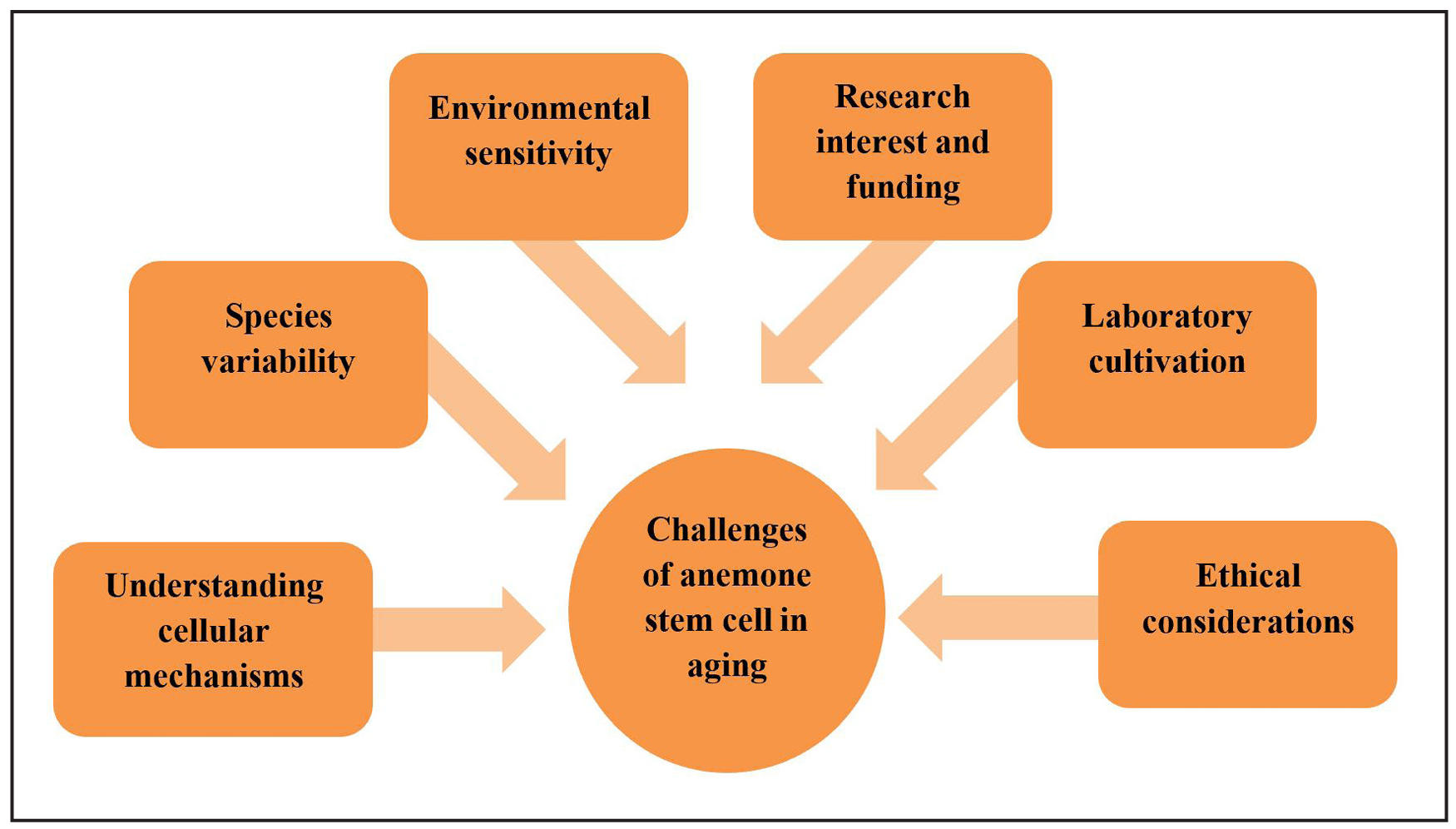
Figure 3. Challenges of anemone stem cell in aging.
Future directions
There are several compelling avenues for future anemone stem cell research, each offering unique insights and applications (Table 4). While the conclusion effectively summarizes the current understanding of aging and regeneration in sea anemones, to advance this field, future studies should focus on several promising areas. Utilizing advanced genomic and transcriptomic tools like RNA-seq and CRISPR-Cas9 gene editing can elucidate the genetic mechanisms underlying regeneration and aging by identifying key regulatory genes [33]. Comparative analyses with other regenerative species such as zebrafish and planarians can reveal conserved and unique pathways, potentially guiding the development of new regenerative therapies [13]. Investigating how anemone stem cells respond to aging-related stress and cellular senescence will deepen our understanding of how to enhance regenerative capacity in older organisms [34]. Additionally, exploring bioactive molecules specific to anemones could inspire biomimetic approaches for treating age-related diseases [48]. Environmental studies examining the effects of pollution, habitat degradation, and climate change on anemone populations are essential, as these factors may influence their regenerative abilities and provide broader ecological insights [41]. Finally, fostering multidisciplinary collaboration among ecologists, molecular biologists, and bioinformaticians will be critical to integrating complex data and fully uncovering the potential of anemone stem cell biology [49]. These future directions will not only deepen our knowledge of anemone biology but also contribute to innovations in regenerative medicine and aging research.
Table 4.
Recent studies on anemone in aging.
| Study title | Key findings | Research | applications | Ref. |
|---|---|---|---|---|
| Identification of mltipotent sem clls in Nematostella vectensis | Discovered multipotent stem cells regulated by nanos and piwi genes, linked to regeneration and negligible aging. | Basis for studying molecular mechanisms of aging and regenerative medicine. | [50] | |
| The sea anemone Nematostella vectensis: mchano-sensitivity, extreme regeneration, and longevity | Demonstrated the organism’s exceptional regenerative abilities and potential as a model for longevity studies. | Useful for biomedical research in aging and regeneration. | [51] | |
| Experimental tools to study regeneration in the sea anemone | Provided methodologies for inducing regeneration and analyzing molecular responses. | Enhances experimental studies on aging and regenerative biology. | [52] | |
| Do sea anemones live forever? | Discussed telomere dynamics contributing to their longevity. | Potential insights into telomere biology and human aging. | [53] | |
| Sea anemone delivery of collagen and γ-PGA for anti-aging benefits | Identified collagen and γ-PGA for potential use in anti-aging skincare. | Applications in cosmetics for youth-preserving formulations. | [54] |
Conclusions
The investigation of aging in sea anemones has produced a number of important discoveries that improve our knowledge of the aging process and the amazing capacity for regeneration exhibited by these creatures. Unique biological traits of anemones include the capacity to restore missing body parts and the presence of multipotent stem cells. These stem cells are essential for maintenance and repair, enabling anemones to heal from wounds and adjust to shifting environmental conditions. However, anemones' ability to regenerate can diminish with age, underscoring the intricate connection between cellular function and aging. Significant biological changes, such as the start of stem cell senescence, modifications to communication networks, and an increase in oxidative stress, have been linked to anemone aging. Further research into the molecular mechanisms underlying these processes is crucial since these factors might collectively hinder regeneration. Gaining knowledge about how anemones control cellular aging and preserve their capacity for regeneration may help us better understand the basic biology of longevity [55, 56].
It is impossible to exaggerate the significance of ongoing study in this area. With potential uses in biogerontology and regenerative medicine, anemones offer a straightforward yet effective model for researching aging and regeneration. Studies of anemones may provide insights into more general biological and medical domains, especially in the areas of aging-related illnesses and the creation of innovative treatment approaches. In conclusion, research on aging in sea anemones advances our knowledge of aging processes in more complicated species, such as humans, while also enhancing our comprehension of these intriguing creatures [57]. We can discover the complex mechanisms governing lifespan and resilience by further investigating the interaction of aging, regeneration, and environmental factors. This will open the door to novel methods to the treatment of health and disease.
Declarations
Authorship Contribution Statement
Gaurav N. Kasar: Conceptualization, Investigation, Writing original draft. Pooja B. Rasal: Conceptualization, Investigation, Writing original draft. Durgesh S. Pagar: Resources, Data curation, Visualization, Formal analysis. Aman B. Upaganlawar: Resources, Data curation, Visualization, Formal analysis. Sunil K. Mahajan: Resources, Data curation, Visualization, Formal analysis
Conflict of interest
The authors confirm that there are no known conflicts of interest.
Availability of data and materials
All data generated or analyzed during this study are included in this published article.
References
1. DiLoreto R, & Murphy C. The cell biology of aging. Mol Biol Cell, 2015, 26(25): 4524-4531. [Crossref]
2. López-Otín C, Blasco M, Partridge L, Serrano M, & Kroemer G. Hallmarks of aging: an expanding universe. Cell, 2023, 186(2): 243-278. [Crossref]
3. Rando T, & Chang H. Aging, rejuvenation, and epigenetic reprogramming: resetting the aging clock. Cell, 2012, 148(1-2): 46-57. [Crossref]
4. Tchkonia T, Zhu Y, van Deursen J, Campisi J, & Kirkland J. Cellular senescence and the senescent secretory phenotype: therapeutic opportunities. J Clin Invest, 2013, 123(3): 966-972. [Crossref]
5. Holstein T. The role of cnidarian developmental biology in unraveling axis formation and Wnt signaling. Dev Biol, 2022, 487: 74-98. [Crossref]
6. Röttinger E. Nematostella vectensis, an emerging model for deciphering the molecular and cellular mechanisms underlying whole-body regeneration. Cells, 2021, 10(10): 2692-2699. [Crossref]
7. Davidson E, Rast J, Oliveri P, Ransick A, Calestani C, Yuh C, et al. A genomic regulatory network for development. Science, 2002, 295(5560): 1669-1678. [Crossref]
8. Layden M, Rentzsch F, & Röttinger E. The rise of the starlet sea anemone Nematostella vectensis as a model system to investigate development and regeneration. Wiley Interdiscip Rev Dev Biol, 2016, 5(4): 408-428. [Crossref]
9. González-Muñoz R, Simões N, Guerra-Castro E, Hernández-Ortíz C, Carrasquel G, Mendez E, et al. Sea anemones (Cnidaria: Actiniaria, Corallimorpharia, Ceriantharia, Zoanthidea) from marine shallow-water environments in Venezuela: new records and an updated inventory. Marine Biodiversity Records, 2016, 9(1): 18-28. [Crossref]
10. van der Burg C, & Prentis P. The tentacular spectacular: evolution of regeneration in sea anemones. Genes, 2021, 12(7): 1072-1082. [Crossref]
11. Lv J, Chen J, Li L, Geng X, Li B, Wang M, et al. Signaling mechanism of budding, proliferation, and tissue regeneration in Cnidaria. Current Issues in Molecular Biology, 2025, 47(4): 219-229.
12. Technau U. Gastrulation and germ layer formation in the sea anemone Nematostella vectensis and other cnidarians. Mechanisms of Development, 2020, 163: 103628. [Crossref]
13. DuBuc T, Dattoli A, Babonis L, Salinas-Saavedra M, Röttinger E, Martindale M, et al. In vivo imaging of Nematostella vectensis embryogenesis and late development using fluorescent probes. BMC Cell Biol, 2014, 15: 44-54. [Crossref]
14. Rentzsch F, Layden M, & Manuel M. The cellular and molecular basis of cnidarian neurogenesis. Wiley Interdiscip Rev Dev Biol, 2017, 6(1): 257-267. [Crossref]
15. Rathbun L, Everett C, & Bergstralh D. Emerging cnidarian models for the study of epithelial polarity. Front Cell Dev Biol, 2022, 10: 854373. [Crossref]
16. Gahan J, Bradshaw B, Flici H, & Frank U. The interstitial stem cells in Hydractinia and their role in regeneration. Curr Opin Genet Dev, 2016, 40: 65-73. [Crossref]
17. Walczyńska K, Zhu L, & Liang Y. Insights into the role of the Wnt signaling pathway in the regeneration of animal model systems. Int J Dev Biol, 2023, 67(3): 65-78. [Crossref]
18. Shi Q, Xue C, Zeng Y, Yuan X, Chu Q, Jiang S, et al. Notch signaling pathway in cancer: from mechanistic insights to targeted therapies. Signal Transduct Target Ther, 2024, 9(1): 128-138. [Crossref]
19. Chen C, McKinney S, Ellington L, & Gibson M. Hedgehog signaling is required for endomesodermal patterning and germ cell development in the sea anemone Nematostella vectensis. Elife, 2020, 9: 54573. [Crossref]
20. Maharati A, & Moghbeli M. PI3K/AKT signaling pathway as a critical regulator of epithelial-mesenchymal transition in colorectal tumor cells. Cell Commun Signal, 2023, 21(1): 201-212. [Crossref]
21. Denner A, Steger J, Ries A, Morozova-Link E, Ritter J, Haas F, et al. Nanos2 marks precursors of somatic lineages and is required for germline formation in the sea anemone Nematostella vectensis. Sci Adv, 2024, 10(33): eado0424. [Crossref]
22. Bosch T. Why polyps regenerate and we don't: towards a cellular and molecular framework for Hydra regeneration. Dev Biol, 2007, 303(2): 421-433. [Crossref]
23. Zhang W, Koyuncu S, & Vilchez D. Insights into the links between proteostasis and aging from C. elegans. Front Aging, 2022, 3: 854157. [Crossref]
24. DuBuc T, Traylor-Knowles N, & Martindale M. Initiating a regenerative response; cellular and molecular features of wound healing in the cnidarian Nematostella vectensis. BMC Biol, 2014, 12: 24-33. [Crossref]
25. Rossiello F, Jurk D, Passos J, & d'Adda di Fagagna F. Telomere dysfunction in ageing and age-related diseases. Nat Cell Biol, 2022, 24(2): 135-147. [Crossref]
26. Campisi J. Aging, cellular senescence, and cancer. Annu Rev Physiol, 2013, 75: 685-705. [Crossref]
27. Kuilman T, Michaloglou C, Vredeveld L, Douma S, van Doorn R, Desmet C, et al. Oncogene-induced senescence relayed by an interleukin-dependent inflammatory network. Cell, 2008, 133(6): 1019-1031. [Crossref]
28. Chocron E, Munkácsy E, & Pickering A. Cause or casualty: the role of mitochondrial DNA in aging and age-associated disease. Biochim Biophys Acta Mol Basis Dis, 2019, 1865(2): 285-297. [Crossref]
29. Singh V, Bali A, Singh N, & Jaggi A. Advanced glycation end products and diabetic complications. Korean J Physiol Pharmacol, 2014, 18(1): 1-14. [Crossref]
30. Salameh Y, Bejaoui Y, & El Hajj N. DNA methylation biomarkers in aging and age-related diseases. Front Genet, 2020, 11: 171-181. [Crossref]
31. Harman D. Aging: a theory based on free radical and radiation chemistry. J Gerontol, 1956, 11(3): 298-300. [Crossref]
32. Anisimova A, Alexandrov A, Makarova N, Gladyshev V, & Dmitriev S. Protein synthesis and quality control in aging. Aging, 2018, 10(12): 4269-4288. [Crossref]
33. Trevino M, Stefanik D, Rodriguez R, Harmon S, & Burton P. Induction of canonical Wnt signaling by alsterpaullone is sufficient for oral tissue fate during regeneration and embryogenesis in Nematostella vectensis. Dev Dyn, 2011, 240(12): 2673-2679. https://doi.org/10.1002/dvdy.22774">Crossref]
34. Petralia R, Mattson M, & Yao P. Aging and longevity in the simplest animals and the quest for immortality. Ageing res rev, 2014, 16: 66-82. [Crossref]
35. Shay J. Role of telomeres and telomerase in aging and cancer. Cancer Discov, 2016, 6(6): 584-593. [Crossref]
36. Melzer D, Pilling L, & Ferrucci L. The genetics of human ageing. Nat Rev Genet, 2020, 21(2): 88-101. [Crossref]
37. Zakrzewski W, Dobrzyński M, Szymonowicz M, & Rybak Z. Stem cells: past, present, and future. Stem Cell Res Ther, 2019, 10(1): 68-78. [Crossref]
38. Reddien P. The cellular and molecular basis for planarian regeneration. Cell, 2018, 175(2): 327-345. [Crossref]
39. Apfeld J, O'Connor G, McDonagh T, DiStefano P, & Curtis R. The AMP-activated protein kinase AAK-2 links energy levels and insulin-like signals to lifespan in C. elegans. Genes Dev, 2004, 18(24): 3004-3009. [Crossref]
40. Hoepner C, Abbott C, & Burke da Silva K. The ecological importance of toxicity: sea anemones maintain toxic defence when bleached. Toxins, 2019, 11(5): 266-276. [Crossref]
41. Hughes T, Kerry J, Álvarez-Noriega M, Álvarez-Romero J, Anderson K, Baird A, et al. Global warming and recurrent mass bleaching of corals. Nature, 2017, 543(7645): 373-377. [Crossref]
42. Zhou K, Pincus Z, & Slack F. Longevity and stress in Caenorhabditis elegans. Aging, 2011, 3(8): 733-753. [Crossref]
43. Gacesa R, Chung R, Dunn S, Weston A, Jaimes-Becerra A, Marques A, et al. Gene duplications are extensive and contribute significantly to the toxic proteome of nematocysts isolated from Acropora digitifera (Cnidaria: Anthozoa: Scleractinia). BMC Genomics, 2015, 16: 774-784. [Crossref]
44. Antelo-Iglesias L, Picallos-Rabina P, Estévez-Souto V, Da Silva-Álvarez S, & Collado M. The role of cellular senescence in tissue repair and regeneration. Mech Ageing Dev, 2021, 198: 111528. [Crossref]
45. Bornstein K, Gryan G, Chang E, Marchler-Bauer A, & Schneider V. The NIH Comparative Genomics Resource: addressing the promises and challenges of comparative genomics on human health. BMC Genomics, 2023, 24(1): 575-585. [Crossref]
46. Chan W, Tay Y, Ang H, Tun K, Chou L, Huang D, et al. Reproduction in urbanised coastal waters: shallow-water sea anemones (Entacmaea quadricolor and Stichodactyla haddoni) maintain high genetic diversity and panmixia. Diversity, 2020, 12(12): 467-478.
47. Mather J. Ethics and invertebrates: the problem is us. Animals, 2023, 13(18): 2827-2837. [Crossref]
48. Atanasov A, Zotchev S, Dirsch V, & Supuran C. Natural products in drug discovery: advances and opportunities. Nat Rev Drug Discov, 2021, 20(3): 200-216. [Crossref]
49. Kaikkonen L, Shellock R, Selim S, Ojwala R, Dias B, Li S, et al. Fostering diversity, equity, and inclusion in interdisciplinary marine science. npj Ocean Sustainability, 2024, 3(1): 49-60. [Crossref]
50. Warner J, Guerlais V, Amiel A, Johnston H, Nedoncelle K, & Röttinger E. NvERTx: a gene expression database to compare embryogenesis and regeneration in the sea anemone Nematostella vectensis. Development, 2018, 145(10): 162867. [Crossref]
51. Amiel A, Michel V, Carvalho J, Shkreli M, Petit C, & Röttinger E. The sea anemone Nematostella vectensis, an emerging model for biomedical research: mechano-sensitivity, extreme regeneration and longevity. Med Sci, 2021, 37(2): 167-177. [Crossref]
52. Amiel A, & Röttinger E. Experimental tools to study regeneration in the sea anemone Nematostella vectensis. Methods Mol Biol, 2021, 2219: 69-80. [Crossref]
53. Ellis M. Do sea anemones live forever? Bay Nature, 2020.
54. Lim Y, Ok Y, Hwang S, Kwak J, & Yoon S. Marine collagen as a promising biomaterial for biomedical applications. Mar Drugs, 2019, 17(8): 467-476. [Crossref]
55. Litsios G, Sims C, Wüest R, Pearman P, Zimmermann N, & Salamin N. Mutualism with sea anemones triggered the adaptive radiation of clownfishes. BMC Evol Biol, 2012, 12: 212-222. [Crossref]
56. Bradshaw B, Thompson K, & Frank U. Distinct mechanisms underlie oral vs aboral regeneration in the cnidarian Hydractinia echinata. Elife, 2015, 4: e05506. [Crossref]
57. Perkins M, Gandara L, & Crocker J. A synthetic synthesis to explore animal evolution and development. Philos Trans R Soc Lond B Biol Sci, 2022, 377(1855): 20200517. [Crossref]