Open Access | Brief
This work is licensed under a Creative Commons Attribution-ShareAlike 4.0 International License.
Voluntary wheel running enhances neuronal BDNF in very old C57BL/6 mice
* Corresponding author: Warren Ladiges
Mailing address: Department of Comparative Medicine, School
of Medicine, University of Washington, Seattle, WA, USA.
Email: wladiges@uw.edu
Received: 13 March 2026 / Revised: 20 March 2026 / Accepted: 26 March 2026 / Published: 30 June 2026
DOI: 10.31491/APT.2026.06.225
Abstract
Aging can lead to cognitive impairment in many individuals, with certain brain regions becoming less active and protein expression decreasing. However, it is possible to slow cognitive decline and preserve brain health with physical activity, which can increase levels of important brain proteins such as brain-derived neurotrophic factor (BDNF). To determine the impact of exercise on various brain proteins, voluntary wheel running (VWR) was observed in aged C57BL/6 mice for 3 days. VWR not only measures muscle performance but also serves as a non-stressful protocol that distinguishes it from forced activity, such as treadmill running or swimming, and more closely mimics natural animal behavior. Following VWR, immunohistochemistry was performed, and tissue scans were analyzed for BDNF, autophagy-related protein 5 (ATG5), and interleukin-6 (IL-6). Expression of these proteins, linked to inflammation, aging, and cell damage, respectively, was assessed in the hippocampus, neocortex, and the hypothalamus. Analysis revealed that VWR increased BDNF expression in the hippocampus and hypothalamus. The effects of VWR on BDNF, ATG5, and IL-6 protein expression differed by brain region and varied between sexes. These results suggest that voluntary exercise may serve as a practical intervention to preserve cognitive function at very old ages.
Keywords
BDNF, voluntary wheel running, aging intervention
Introduction
Aging is a complex biological process often accompanied by cognitive decline and increased susceptibility to
neurodegenerative diseases. Current therapeutic interventions targeting age-related cognitive impairment remain
limited in efficacy. As a result, there is growing interest in
identifying non-pharmacological strategies, like exercise,
that may help preserve brain health and delay the onset of
neurological dysfunction.
Voluntary wheel running (VWR) is a model of physical
activity that closely resembles natural behavior, making
it a valuable tool for studying the effects of exercise on
the aging brain. VWR minimizes stress and better reflects natural mouse behavior, giving better insight into how lifestyle factors influence brain function. Additionally, VWR closely mirrors everyday human physical activity. Therefore, these findings can help guide the use of exercise as a practical approach to support brain health during aging and to develop biomarker-guided interventions for older populations.
The aim of this study was to investigate how short-term VWR affects inflammation, autophagy, and neuronal health through marker analysis. The markers examined were IL-6, ATG5, and BDNF in the brains of very old C57BL/6 mice. In addition, cresyl violet staining, which marks nissl substances in the endoplasmic reticulum (ER) of neurons, was used to assess neuronal health and structure. The marker analysis would ultimately reveal whether voluntary exercise could mitigate age-related molecular changes and potentially serve as a preventative measure against cognitive decline.
This study focuses on three key brain proteins with significant roles in aging and neural health. Brain-derived neurotrophic factor (BDNF) supports synaptic plasticity and neurogenesis, and its activity decreases in the aging brain [1]. Additionally, interleukin-6 (IL-6), a
pro-inflammatory cytokine, is associated with neuroinflammation and cognitive impairment [2]. ATG5, an autophagy-related protein, is essential for cellular homeostasis and it declines with age. A lack of functional ATG5 means the cell cannot dispose of its damaged components [3].
Protein expression was analyzed in the hippocampus, hypothalamus, and neocortex following immunohistochemistry (IHC). These brain regions are all heavily involved in learning, memory, and higher-level thinking, and they are also extremely vulnerable to aging. The hippocampus plays a central role in memory and learning; abilities that become much more difficult with age [4]. The hypothalamus is critical for homeostasis and hormone regulation and influences appetite and thirst, as well as sexual behavior. The hypothalamus is known to become less active with age. The neocortex is mainly connected with higher order brain functions such as learning, reasoning, and problem-solving [5]. These regions reveal overall cognitive health and studying them will allow for a better understanding of how voluntary exercise might counteract molecular changes that underlie cognitive decline. Ultimately, these regions offer insight into potential treatments for age-related cognitive disorders.
Methods
Animals and experimental design
30-month male (n = 7) and 32-month female (n = 9) C57BL/6 mice from the NIA Aged Mouse Colony (Charles River, Inc.) were used in this experiment. Mice were given access to running wheels (Med. Associates, Inc.) for a duration of three days, as previously described [6]. They were able to run freely on the open surface of a slanted plastic saucer-shaped wheel placed inside of a standard mouse cage. The running wheel had a diameter of 15.5 cm, corresponding to a running distance of approximately 49 cm per full rotation. Rotations were transmitted electronically to a USB hub so that the rate of running and frequency could be captured to a software program for analysis and data storage. Each mouse was housed and observed individually. Furthermore, all mice were subject to the same standard cages with identical bedding, food, and water conditions. Before the running assessment began, wheels were locked for 48 hours so the mice could get used to the wheel, thereby reducing behavioral disturbances. Two male mice and three female mice had locked wheels and were assigned as the control group.
IHC
Following the experiment, mice were euthanized using carbon dioxide as the primary method according to American Veterinary Medical Association (AVMA) guidelines. Brains were collected and fixed in formalin for 48 hours before being switched to PBS for storage. Tissues were paraffin-embedded and sectioned into four-micrometer slides for IHC staining. IHC was performed according to manufacturer protocol (Abcam HRP/DAB Rabbit Kit: ab64261). The slides were rehydrated using graded ethanol and xylene, followed by heat-mediated antigen retrieval at 98 °C in citric acid buffer (pH 6.0). Endogenous peroxidase activity as well as non-specific binding was blocked using serum, avidin, and biotin. Slides were incubated overnight at 4 °C with primary antibodies targeting BDNF (Abcam ab213323 at 1:200) and ATG5 (ThermoFisher MA5-35339 at 1:500). Slides were treated with a biotinylated secondary antibody and streptavidin-HRP, then developed using DAB chromogen. Finally, slides were dehydrated and mounted before photographing.
Cresyl violet staining
An additional set of slides was stained with cresyl violet (Abcam ab246817). These slides underwent the same rehydration steps as used in IHC, followed by 5 minutes in cresyl violet solution. Excess stain was removed with three rinses in 100% ethanol, followed by 3-minutes in xylene. Slides were then coverslipped for analysis.
Digital imaging
Photos were taken at 4x and 20x on a microscope using NIS Elements for image analysis. The ImageJ extension through QuPath (version Mac OS X 10.16) was used to evaluate the percent of positive staining for ATG5 and IL-6 in the hippocampus and for BDNF in the hippocampus, hypothalamus, and the neocortex.
Statistical analysis
All statistical analyses were performed and analyzed using GraphPad Prism. T-tests were used to compare the control and running groups for percent positive staining of BDNF and ATG5 in the hippocampus. A significance level of 0.05 was used.
Results and discussion
VWR produced a distance-dependent increase in BDNF expression, with mice covering greater distances (running farther) showing higher BDNF levels (Figure 1). The data suggested a positive association between BDNF expression and the distance covered by the mice, indicating that higher physical activity is linked to increased neurotrophic support.
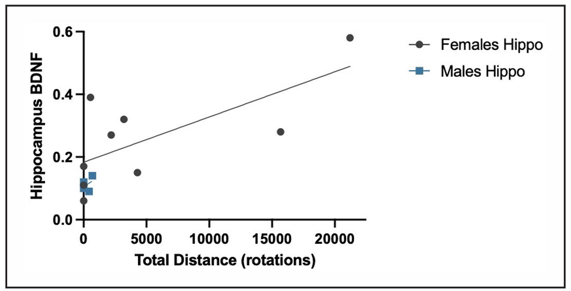
Figure 1. Distance ran compared to BDNF. The distance ran was compared to the percentage of positive staining of hippocampal BDNF. Mice that ran farther tended to show higher percentages of positive BDNF staining, indicating a distance-related pattern in BDNF expression.
Region-specific analysis revealed that the VWR group had a significantly higher average of BDNF staining than the control group in the hippocampus and hypothalamus but not in the neocortex (Figure 2). In the hippocampus, females exhibited a significant increase (P = 0.0438), whereas males showed no significant effect (P = 0.87), suggesting a sex-specific response. However, the smaller sample size in this group may have limited the ability to detect the true effect. For females, since the P-value of 0.0438 < 0.05, we can reject the null hypothesis, therefore, we can conclude that VWR had a statistically significant effect on hippocampal BDNF levels. For males, VWR does not have a statistically significant effect on hippocampal BDNF levels.
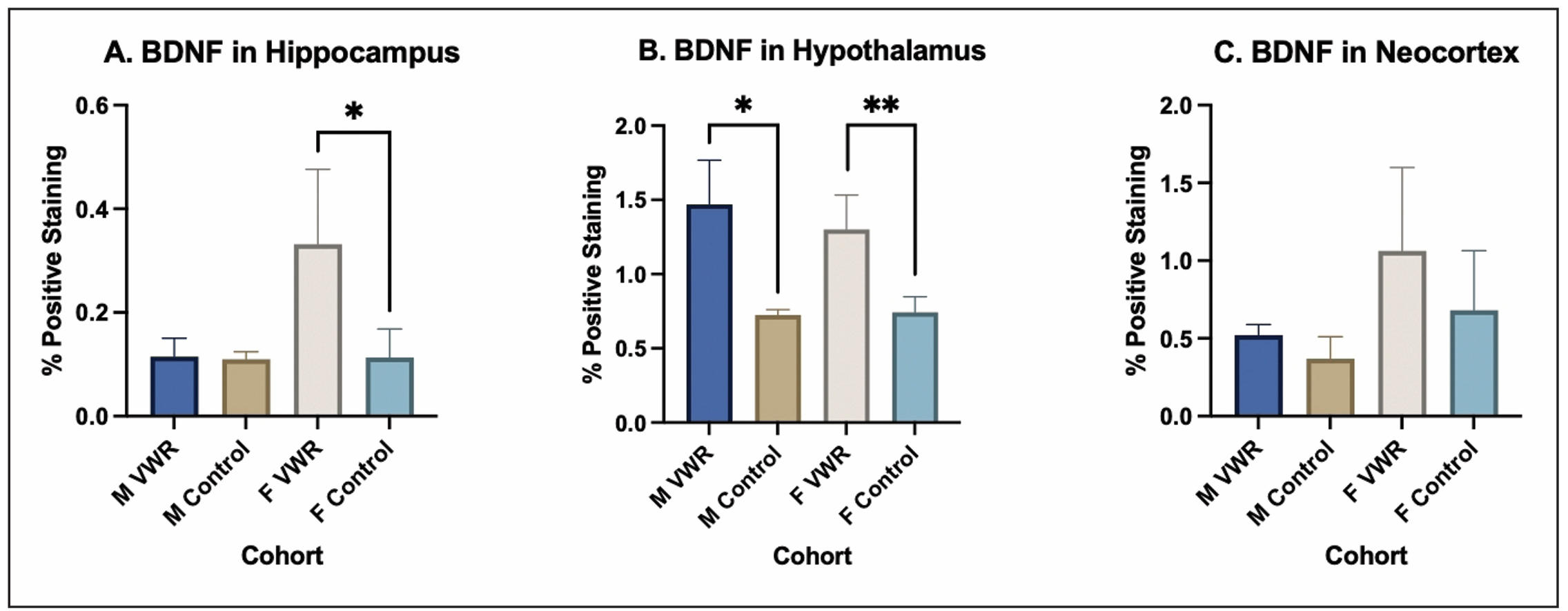
Figure 2. Region-specific BDNF expression in aged mice following VWR. (A) Hippocampal BDNF expression was higher in the VWR group compared to sedentary controls. (B) BDNF levels in the hypothalamus were elevated following VWR. (C) No significant differences were observed in neocortical BDNF between groups.
In the hypothalamus, both males (P = 0.0444) and females (P = 0.0063) demonstrated significant increases, with females showing a slightly stronger effect. For females, since the P-value of 0.0063 < 0.05, we can reject the null hypothesis. Therefore, we can conclude that voluntary wheel running had a statistically significant effect on hippocampal BDNF levels in females. In addition, physical activity had a significant effect on BDNF levels in males (P = 0.0444). Since the P-value of 0.0444 < 0.05, we can reject the null hypothesis. Thus, physical activity did have a statistically significant effect on BDNF levels in the hypothalamus.
As for BDNF levels in the neocortex, the P-value for both males and females exceeded 0.05, thus voluntary wheel running had no significant effect on BDNF levels in the neocortex and we fail to reject the null hypothesis. The absence of changes in neocortical BDNF highlights that exercise-induced neurotrophic enhancement is regionally selective.
Markers of neuronal health, including neuronal count (cresyl violet), inflammation (IL-6), and autophagy (ATG5) were examined in the hippocampus (Figure 3). These markers were unaffected by VWR (P > 0.05), indicating that voluntary running did not alter these specific measures in very old mice. These results show that the observed increases in BDNF occurred
alongside stable levels of these hippocampal markers. Since the P-values for both males and females exceeded 0.05 we must fail to reject the null and can conclude VWR had no significant effect on IL-6, ATG5, or CV levels. These results indicate that voluntary wheel running does not induce brain inflammation or other adverse effects, suggesting that exercise is safe and does not negatively impact neuronal health or overall condition in the animals.
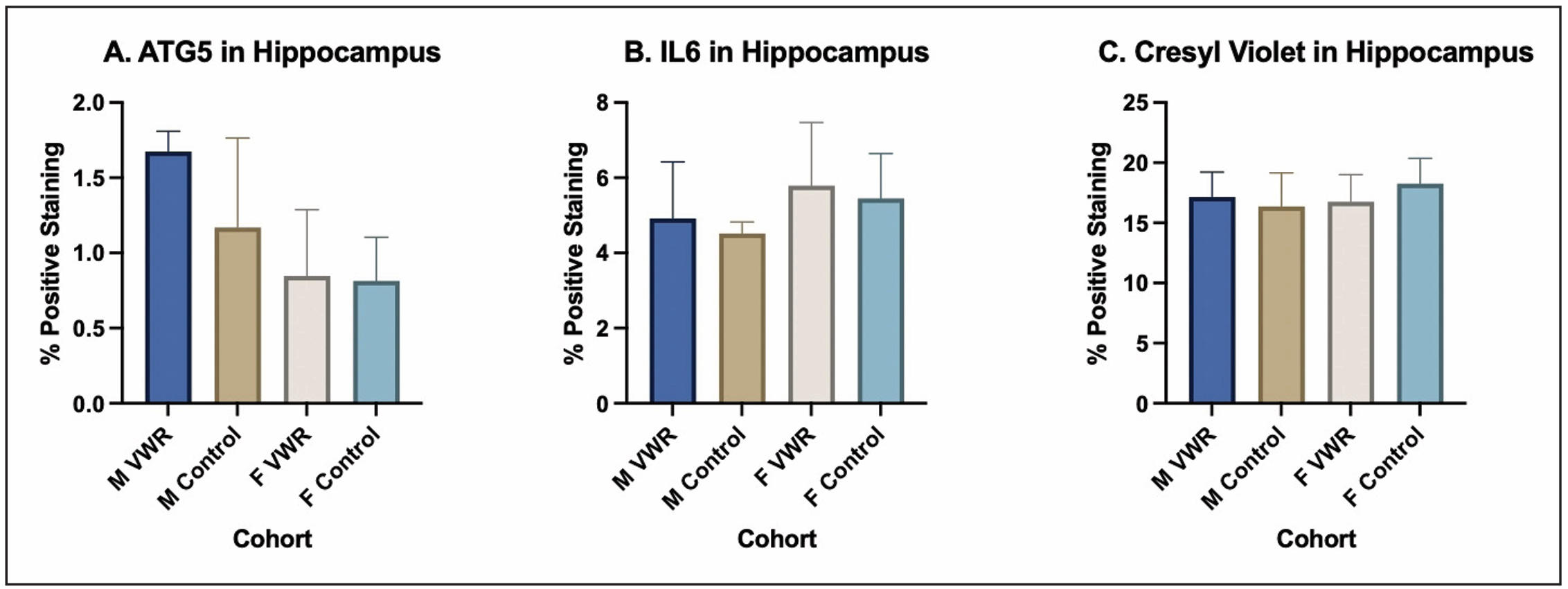
Figure 3. Hippocampal brain health markers in aged mice following VWR. (A) ATG5 expression was measured as a marker for autophagy. (B) IL-6 expression was assessed as a marker for inflammation. (C) Cresyl Violet staining was used to determine neuronal counts in the hippocampus. No significant differences were observed in ATG5, IL-6, or Cresyl Violet staining following VWR, suggesting that these pathways were not detectably altered by voluntary wheel running in very old mice.
Collectively, these results indicate that VWR in very old mice—an age rarely examined in exercise studies—is associated with region-specific changes in hippocampal and hypothalamic BDNF expression, with sex differences especially evident in hippocampal responses. No significant changes were observed in hippocampal markers of neuronal count (cresyl violet), inflammation (IL-6), or autophagy (ATG5), indicating that these BDNF changes occurred without detectable alterations in these specific measures of neuronal health. While statistically significant, the practical importance and biological relevance of these results may be influenced by differences in strain, age, sex and individual motivation. The small sample size could also influence the strength of these conclusions. Future studies incorporating standardized behavioral assessments and quantitative analyses are warranted to clarify how increased BDNF triggered by physical exercise affects brain function in geriatric mice, which will help provide additional insights into gerotherapeutic intervention of aging in older people.
Declarations
Availability of data and materials
Not applicable.
Financial support and sponsorship
None.
Conflicts of interest
Warren Ladiges is a member of the editorial board of Aging Pathobiology and Therapeutics. The authors declare that they have no conflicts and were not involved in the journal’s review or decision regarding this manuscript.
Consent for publication
Not applicable.
AI and AI-assisted tools statement
No AI tools were used in the preparation of this article.
References
1. Colucci-D'Amato L, Speranza L, & Volpicelli F. Neurotrophic factor BDNF, physiological functions and therapeutic potential in depression, neurodegeneration and brain cancer. Int J Mol Sci, 2020, 21(20): 7777. [Crossref]
2. Aliyu M, Zohora F, Anka A, Ali K, Maleknia S, Saffarioun M, et al. Interleukin-6 cytokine: An overview of the immune regulation, immune dysregulation, and therapeutic approach. Int Immunopharmacol, 2022, 111: 109130. [Crossref]
3. Changotra H, Kaur S, Yadav S, Gupta G, Parkash J, & Duseja A. ATG5: a central autophagy regulator implicated in various human diseases. Cell Biochem Funct, 2022, 40(7): 650-667. [Crossref]
4. Fogwe L, Reddy V, Mesfin F. Neuroanatomy, hippocampus. Treasure Island (FL): StatPearls Publishing, 2023.
5. Borrell V. Recent advances in understanding neocortical development. F1000Res, 2019, 8(1791): 20332.1. [Crossref]
6. Goh J, & Ladiges W. Voluntary wheel running in mice. Curr Protoc Mouse Biol, 2015, 5(4): 283-290. [Crossref]