Open Access | Case Report
This work is licensed under a Creative Commons Attribution-ShareAlike 4.0 International License.
Wandering spleen with torsion presenting as a rare case of acute abdomen
Corresponding author: Solomon Bekele Abeb
Mailing address: Department of Surgery, Addis Ababa University, College of Health Sciences, Addis Ababa, Ethiopia.
E-mail: solbekeleabebe@gmail.com
Received: 13 May 2021 / Accepted: 08 September 2021
DOI: 10.31491/CSRC.2021.12.083
Abstract
Wandering spleen is a rare clinical occurrence characterized by the absence of spleen in its normal anatomic place. Patients may present with acute abdomen, abdominal mass, and chronic abdominal pain. Prompt diagnosis and intervention are necessary. Here, we report a case of a woman who presented with acute abdominal pain secondary to a wandering spleen complicated by torsion of its vascular pedicle.
Keywords
Wandering spleen; torsion; splenectomy
Introduction
The spleen develops from the mesoderm in the dorsal
mesogastrium. It lies in the left hypochondrium behind
the stomach, and is approximately 12 cm long and 7
cm wide. The spleen is fixed in position by the lienorenal and gastrosplenic ligaments; the phrenicocolic
ligament provides additional support. The ligaments
are embryological condensations that take place in the
peritoneum, and congenital peritoneal anomalies may
result in splenic displacement [1, 2].
Wandering spleen, also known as displaced, ectopic,
drifting, floating spleen, or Solenopsis is a rare condition defined as a huge, single spleen in an abdominal
position rather than its anatomical site, owing to laxity
of its pedicles and absence of ligamentous attachments
[3].
Acquired anomalies have been described and are attributed to laxity of the ligaments due to weakness of
the abdominal wall, multiple pregnancies, hormonal
changes, or an increase in size in the spleen. Both congenital and acquired conditions result in a long pedicle,
which is predisposed to torsion. The splenic vessel’s
Case Report
course within the pedicle, and therefore, torsion of
the pedicle results in partial or complete infarct of the
spleen [3, 4].
We report a case of torsion of a wandering spleen in a
40 years old multiparous woman who presented to our
emergency with an acute abdomen.
Case Report
A 40 years old female patient presented to our emergency room with abdominal pain and progressive
abdominal distention of a two-week duration which
got worsen over the past 24 hours. She claims to have
3 episodes of vomiting of ingested matter. She had no
prior similar illness nor chronic medical conditions.
She is a para 8 mother from rural Ethiopia. Physical examination revealed tachycardia of 100/min.
Abdominal examination showed a huge, firm, tender
periumbilical mass measuring 12 x 8 cm extending to
the right lower quadrant area, with positive signs of
fluid collection. Abdominal radiography showed an
important colon distention especially at the upper left
quadrant without air-fluid levels (Figure 1). Abdominal
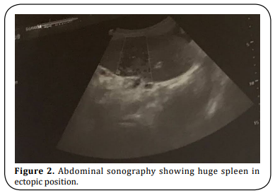
ultrasound is done showed a midline pelvic region solid mass, measuring 12.8 x 6.3 x 14.9 cm in size (Figure
2). There were multiple internal echogenic foci with no
distal shadowing, likely micro hemorrhage, and spleen
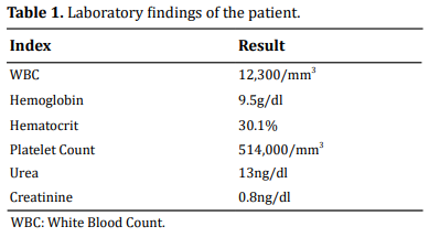
not visualized in its normal anatomic location. Laboratory findings of the patient are shown in Table 1.
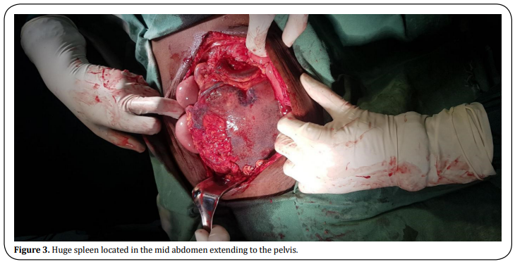
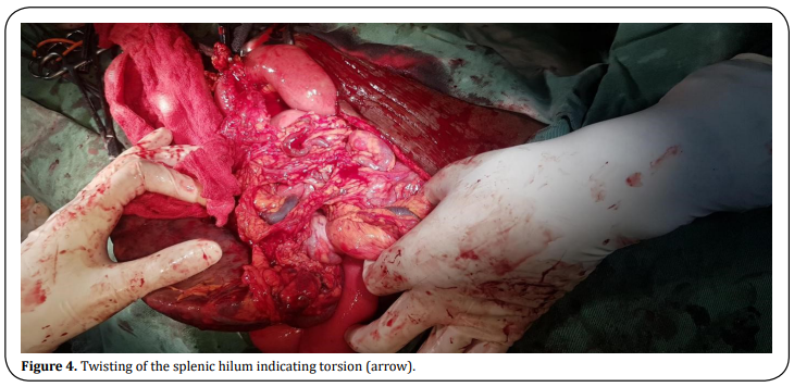
Splenectomy was performed avoiding injury to the pancreatic tail (Figure 3-5). The histology section showed tissue mainly composed of hemorrhage, congested blood vessels with lymphocyte, plasma cells, and neutrophils. The features were consistent with splenic infarction of ischemic origin. The postoperative period was uneventful. The patient was discharged on the fourth postoperative day, to come back for vaccination two weeks later.


Discussion
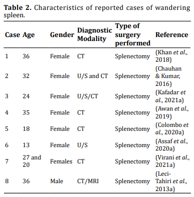
Wandering spleen is a rare clinical finding where the spleen is found suspended by only its mesentery. It is usually found in young children and women aged between 20 and 40 years of age. Most cases are attributed to be congenital in origin but acquired conditions like multiple pregnancies have also been incriminated. The splenic vessel’s course within the pedicle, and therefore, torsion of the pedicle results in a partial or complete infarct of the spleen [5]. Torsion of a wandering spleen is diagnosed in about 0.2 - 0.3% of patients who require splenectomy. The clinical presentation of the wandering spleen may be variable, from an asymptomatic patient to one with mild abdominal pain, signs of acute abdomen with or without peritonitis [2, 6]. Ultrasound imaging with duplex scanning can be used as an initial mode of imaging which can show the position of the wandering spleen with concomitant replacement of bowel in the left upper quadrant. CT contrast imaging is the preferred mode of investigation, with the contrast helping to elucidate the viability of the spleen. An early diagnosis and surgical care are the best guarantees for preserving the spleen [7] (Table 2).
Conclusion
A wandering spleen must be suspected in patients who present with acute abdomen and a palpable mass. Diagnosis requires a high index of clinical suspicion. Abdominal ultrasound and CT are important adjuncts to diagnosis. Splenopexy or splenectomy are options of management depending on whether or not there is infarction.
Declarations
Authors’ contributions
Solomon involved in pre-operative preparation, operation, post op follow-up and wrote this article. Henok supervised the surgery and checked the article. Yonas assisted in the surgery and followed patient postoperatively.
Conflicts of interest
The authors declare that there are no conflicts of interest to disclose.
Ethical approval and consent to participate
The study, which included human samples, complied with the Declaration of Helsinki and obtained written informed consent from all patients involved.
References
1. Chauhan, N. S., & Kumar, S. (2016). Torsion of a
Wandering Spleen Presenting as Acute Abdomen.
Polish Journal of Radiology, 81, 110-113.
2. Khan, D. B., Khandwala, K., Abbasi, S. U., Khan, S.
D., & Raza, R. (2018). Torsion of Wandering Spleen
with Infarction. Cureus, 10(8), e3177.
3. Kafadar, M. T., Daduk, Y., & Karakoç, M. (2021).
Acute abdomen due to torsion of the wandering
spleen: A rare clinical presentation. Ulusal Travma
Veacil Cerrahi Dergisi, 27(1), 154-156.
4. Assaf, R., Shebli, B., Alzahran, A., Rahmeh, A. R.,
Mansour, A., Hamza, R., ... & Ayoub, K. (2020). Acute
abdomen due to an infarction of wandering spleen:
case report. Journal of Surgical Case Reports,
2020(2), rjz378.
5. Virani, P., Farbod, A., Niknam, S., & Akhgari, A.
(2021). Wandering spleen with splenic torsion: Report of two cases. International Journal of Surgery
Case Reports, 78, 274-277.
6. Colombo, F., D’Amore, P., Crespi, M., Sampietro, G.,
& Foschi, D. (2020). Torsion of wandering spleen
involving the pancreatic tail. Annals of Medicine
and Surgery, 50, 10-13.
7. Awan, M., Gallego, J. L., Al Hamadi, A., & Vinod, V.
C. (2019). Torsion of wandering spleen treated by
laparoscopic splenopexy: A case report.International Journal of Surgery Case Reports, 62, 58-61.
8. Leci-Tahiri, L., Tahiri, A., Bajrami, R., & Maxhuni, M.
(2013). Acute abdomen due to torsion of the wandering spleen in a patient with Marfan Syndrome.
World Journal of Emergency Surgery, 8(1), 30.